This is your brain on recovery: A look at the brain over time during abstinence after alcohol use disorder.
This study investigated how the brain recovers from alcohol use disorder during prolonged abstinence, as well as what factors might influence this process. By providing a glimpse into the brain’s ability to regenerate and repair alcohol-related damage, this study offers vital clues for enhancing brain-based treatment approaches for alcohol use disorder.
Long-term, heavy alcohol use can result in a thinning of the brain’s cortex, the outer layer of the brain responsible for many key functions, including decision-making, emotion regulation, and self-control. This cortical thinning process not only constitutes alcohol-related brain damage, but also can translate to potential difficulties for affected individuals in aspects such as critical thinking and social interactions. The concern, especially for those whose drinking meets the criteria for severe alcohol use disorder, is that these challenges could persist or worsen over time, even after the individual stops drinking.
Some research has shown that this cortical thinning can be reversed through short-term abstinence (6 months or less). However, the extent to which these improvements continue over the long term (beyond 6 months), and whether the brain can fully return to a ‘healthy’ state, remains unknown. These questions are especially relevant for individuals with alcohol use disorder, who also suffer from other health conditions that commonly accompany heavy drinking, such as heart disease or diabetes.
To address these questions, this study examined the thickness of the cortex over a period of approximately 7 months among abstinent individuals with a history of alcohol use disorder following outpatient treatment. The study aimed to map the trajectory of the brain’s recovery, looking at how quickly and extensively the alcohol-related cortical thinning can be reversed and whether factors like additional health conditions and smoking affect this recovery. Studies like this one may help illuminate the brain’s potential to recover from the effects of alcohol, thereby informing treatment strategies and support systems for individuals in or seeking recovery from alcohol use disorder.
HOW WAS THIS STUDY CONDUCTED?
Participants with alcohol use disorder were enrolled from treatment programs at two sites: the San Francisco VA Medical Center and Kaiser Permanente outpatient clinics. The control group, individuals without alcohol use disorder, were recruited from the general Bay Area population. The study initially had 68 alcohol use disorder participants; however, some had a recurrence of alcohol or other drug use, or were excluded due to data quality issues, leaving 40 who maintained abstinence until the final assessment.
Researchers used magnetic resonance imaging (MRI) to measure cortical thickness in the brain, comparing alcohol use disorder participants to a control group without alcohol use disorder and who were non-smokers. This comparison helped gauge normal variation in cortical thickness. Both groups were scanned at the study’s onset, and controls were scanned again after 9.6 months. The analysis covered 34 brain regions, and a specialized software was employed to process this structural brain data.
The analysis included both cross-sectional and longitudinal components. In the cross-sectional analysis, the groups were compared at a single point in time. The alcohol use disorder group was further categorized by the presence or absence of heart-related health conditions, which are risk factors for heart disease.
For the longitudinal analysis, the focus was on the alcohol use disorder group over 7.3 months of abstinence, tracking changes in cortical thickness. The analysis took into account age, brain size, average alcohol consumption, days of abstinence, and other factors such as heart-related health conditions, smoking status, and co-occurring anxiety or mood psychiatric disorders (either current or lifetime were considered “positive” for co-occurring mental health disorder). . A specific analysis for alcohol use disorder participants who smoked examined the relationship between the extent of smoking (measured in pack-years, a metric in smoking research combining cigarettes per day and length of time over which someone smoked) and changes in cortical thickness. This was to determine if smoking intensity affected brain recovery.
The final step compared cortical thickness changes in the alcohol use disorder group with those in the control group from their initial to final scans. Adjustments were made for group attributes, brain size, age, and time between scans. This comparison was intended to identify if recovery patterns in alcohol use disorder participants differed from those without alcohol use disorder.
The alcohol use disorder group, recruited primarily from a VA setting, was 51 years old on average, 90% Male, and 80% White. They averaged 13 drinks per day in the year before outpatient treatment. One in 5 had a co-occurring substance use disorder in addition to alcohol, 50% a psychiatric disorder, and 65% had history of hypertension.
WHAT DID THIS STUDY FIND?
Significant recovery in cortical thickness observed, most changes occur early on.
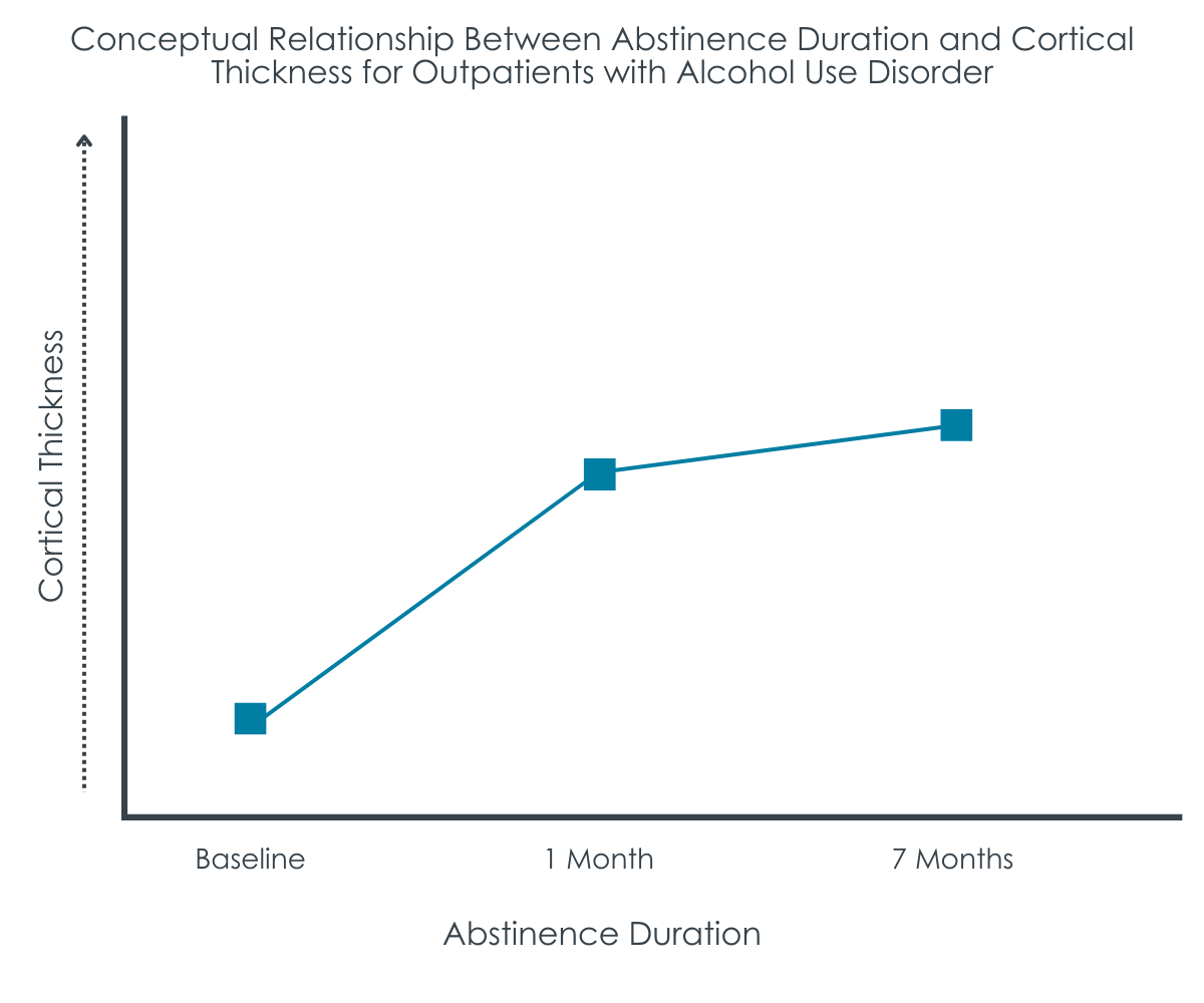
Over the course of 7 months of abstinence, individuals with alcohol use disorder experienced significant improvements in their brain structure. In 25 out of 34 brain regions, the cortex became thicker.
This increase in thickness was not consistent over time; it was more rapid between the 1-week and 1-month marks than between the 1-month mark and the end of the study period.
By the end of the study, the cortical thickness in people with alcohol use disorder was nearly the same as that of the control group in 24 of 34 brain regions examined, indicating a return to a more typical brain structure after prolonged abstinence.
Health conditions and smoking hinder cortical recovery.
The study found that participants with alcohol use disorder who also had heart-health conditions had persistently thinner areas in certain regions of the brain. Moreover, among those who smoked, heavier smoking was associated with less cortical recovery, particularly in the anterior frontal regions of the brain, emphasizing the impact of smoking on brain recovery.
WHAT ARE THE IMPLICATIONS OF THE STUDY FINDINGS?
This study sheds light on how the brain heals in individuals with alcohol use disorder who stop drinking, highlighting that brain recovery is influenced by several factors, including age, alcohol consumption history, overall health, and smoking habits. The findings of this study suggest that the brain has the capacity for physical recovery with continued abstinence, demonstrated by increased thickness in certain brain regions after 7.3 months of abstinence. This supports the idea that some of the neurological effects of chronic, heavy alcohol use may be at least partially reversible and underscores the importance of maintaining abstinence. The degree to which these benefits would have been observed with the elimination of heavy drinking days, rather than all drinking and other drug use days, is unclear, and an important area for future research.
The increase in brain thickness was more pronounced during early abstinence compared to the extended phase. This finding suggests that the brain’s recovery from the effects of alcohol may occur more rapidly at the beginning, with the rate of recovery slowing down over time. The study also highlights that health conditions related to arterial plaque buildup impeded brain recovery in this sample of abstinent individuals who received treatment for alcohol use disorder, pointing to the importance of cardiovascular health in the recovery process. The absence of additional negative impacts from smoking and co-occurring psychiatric or substance use disorders on cortical thickness recovery offers some reassurance that these factors may not impede the brain’s structural recovery capacity during abstinence.
Furthermore, the amount of alcohol consumed before the study was found to be negatively associated with brain recovery. This finding underscores that while the brain can reverse some of the harms experienced with long-term heavy drinking, these toxicity-related impacts persist to some degree, pointing to the clinical benefits of early intervention and relapse prevention among those who enter remission. Overall, however, after 7 months of abstinence, the alcohol use disorder group’s brain thickness in most regions was largely similar to that of the control group, suggesting that substantial cortical recovery is possible.
The study’s conclusions are based on a relatively small group of participants who remained abstinent after outpatient treatment and completed the 7.3-month follow-up. This small sample size means we should be cautious about applying these results to all individuals recovering from alcohol use disorder. Also, with a small percentage of female participants, the study’s findings may not fully represent the recovery process in women with alcohol use disorder, limiting the understanding of gender-specific recovery patterns.
The research did not separately analyze the effects of individual health conditions like high blood pressure or diabetes on brain recovery, rather aggregating them into a single category of “atherogenic” conditions, potentially missing nuances in how these conditions might impact healing. Similarly, the study grouped various psychiatric conditions into one broad category, which may not accurately reflect the distinct impact these conditions have on brain structure.
Apart from smoking, the study did not account for other lifestyle and health factors that could influence brain health, such as physical activity levels, respiratory and liver health, sleep quality, and genetic background.
BOTTOM LINE
This investigation illuminates the dynamic nature of brain recovery in alcohol use disorder, revealing a substantial restoration of cortical thickness over 7 months of abstinence. It also spotlights the importance of addressing vascular health and smoking cessation as part of comprehensive recovery strategies for individuals with alcohol use disorder.
For individuals and families seeking recovery: Findings showed that brain health begins to improve notably within the first month of abstaining from alcohol, indicating a significant period of healing. However, this improvement appears to be lessened for those who smoke or have certain health issues like high blood pressure or diabetes. While the study did not directly link these changes to improvements in day-to-day functioning, the results suggest a positive effect of abstinence on brain health. The key takeaway is that maintaining abstinence from alcohol could lead to meaningful benefits for brain structure and function.
For treatment professionals and treatment systems: The findings from this longitudinal study offer encouraging evidence that substantial cortical recovery is possible in individuals with alcohol use disorder during periods of abstinence, with the most notable changes occurring in the first month. These insights should reinforce the value of early and consistent abstinence in patient care strategies. The study also indicates that this neurological improvement may be weaker for patients who are smokers or have concurrent health conditions. An integrated approach to treatment, addressing both alcohol use and these additional health concerns, including smoking cessation, may enhance recovery outcomes.
For scientists: This study underscores the brain’s capacity for neuroplasticity during the initial phase of abstinence in individuals with alcohol use disorder, with a noted decrease in recovery rate associated with smoking and conditions that affect cardiovascular health. The variable rates of recovery observed across different brain areas are consistent with theories of neuroadaptation, which suggest a period of rapid recovery following cessation of alcohol intake. The study had key methodological limitations, such as a small participant cohort in the later stages of the study, limited gender representation, and other uncontrolled factors, which may impact the interpretation and generalizability of the results. Future research should incorporate a broader range of biopsychosocial factors and longitudinally assess the relationship between structural brain changes and cognitive function during recovery from alcohol use disorder.
For policy makers: The study offers evidence that abstinence from alcohol can lead to substantial improvements in brain structures associated with critical thinking and emotion regulation, with the bulk of this recovery occurring within the first month. These findings highlight the potential impact of early and sustained abstinence on brain health. Policymakers should consider funding integrated treatment programs that address both alcohol use disorder and concurrent medical conditions, such as cardiovascular health issues. The study’s limitations, including the need for more diverse research populations, call for increased funding to support comprehensive research with broad applications, which is critical to informing future healthcare policy.
Long-term, heavy alcohol use can result in a thinning of the brain’s cortex, the outer layer of the brain responsible for many key functions, including decision-making, emotion regulation, and self-control. This cortical thinning process not only constitutes alcohol-related brain damage, but also can translate to potential difficulties for affected individuals in aspects such as critical thinking and social interactions. The concern, especially for those whose drinking meets the criteria for severe alcohol use disorder, is that these challenges could persist or worsen over time, even after the individual stops drinking.
Some research has shown that this cortical thinning can be reversed through short-term abstinence (6 months or less). However, the extent to which these improvements continue over the long term (beyond 6 months), and whether the brain can fully return to a ‘healthy’ state, remains unknown. These questions are especially relevant for individuals with alcohol use disorder, who also suffer from other health conditions that commonly accompany heavy drinking, such as heart disease or diabetes.
To address these questions, this study examined the thickness of the cortex over a period of approximately 7 months among abstinent individuals with a history of alcohol use disorder following outpatient treatment. The study aimed to map the trajectory of the brain’s recovery, looking at how quickly and extensively the alcohol-related cortical thinning can be reversed and whether factors like additional health conditions and smoking affect this recovery. Studies like this one may help illuminate the brain’s potential to recover from the effects of alcohol, thereby informing treatment strategies and support systems for individuals in or seeking recovery from alcohol use disorder.
HOW WAS THIS STUDY CONDUCTED?
Participants with alcohol use disorder were enrolled from treatment programs at two sites: the San Francisco VA Medical Center and Kaiser Permanente outpatient clinics. The control group, individuals without alcohol use disorder, were recruited from the general Bay Area population. The study initially had 68 alcohol use disorder participants; however, some had a recurrence of alcohol or other drug use, or were excluded due to data quality issues, leaving 40 who maintained abstinence until the final assessment.
Researchers used magnetic resonance imaging (MRI) to measure cortical thickness in the brain, comparing alcohol use disorder participants to a control group without alcohol use disorder and who were non-smokers. This comparison helped gauge normal variation in cortical thickness. Both groups were scanned at the study’s onset, and controls were scanned again after 9.6 months. The analysis covered 34 brain regions, and a specialized software was employed to process this structural brain data.
The analysis included both cross-sectional and longitudinal components. In the cross-sectional analysis, the groups were compared at a single point in time. The alcohol use disorder group was further categorized by the presence or absence of heart-related health conditions, which are risk factors for heart disease.
For the longitudinal analysis, the focus was on the alcohol use disorder group over 7.3 months of abstinence, tracking changes in cortical thickness. The analysis took into account age, brain size, average alcohol consumption, days of abstinence, and other factors such as heart-related health conditions, smoking status, and co-occurring anxiety or mood psychiatric disorders (either current or lifetime were considered “positive” for co-occurring mental health disorder). . A specific analysis for alcohol use disorder participants who smoked examined the relationship between the extent of smoking (measured in pack-years, a metric in smoking research combining cigarettes per day and length of time over which someone smoked) and changes in cortical thickness. This was to determine if smoking intensity affected brain recovery.
The final step compared cortical thickness changes in the alcohol use disorder group with those in the control group from their initial to final scans. Adjustments were made for group attributes, brain size, age, and time between scans. This comparison was intended to identify if recovery patterns in alcohol use disorder participants differed from those without alcohol use disorder.
The alcohol use disorder group, recruited primarily from a VA setting, was 51 years old on average, 90% Male, and 80% White. They averaged 13 drinks per day in the year before outpatient treatment. One in 5 had a co-occurring substance use disorder in addition to alcohol, 50% a psychiatric disorder, and 65% had history of hypertension.
WHAT DID THIS STUDY FIND?
Significant recovery in cortical thickness observed, most changes occur early on.
Over the course of 7 months of abstinence, individuals with alcohol use disorder experienced significant improvements in their brain structure. In 25 out of 34 brain regions, the cortex became thicker.
This increase in thickness was not consistent over time; it was more rapid between the 1-week and 1-month marks than between the 1-month mark and the end of the study period.
By the end of the study, the cortical thickness in people with alcohol use disorder was nearly the same as that of the control group in 24 of 34 brain regions examined, indicating a return to a more typical brain structure after prolonged abstinence.
Health conditions and smoking hinder cortical recovery.
The study found that participants with alcohol use disorder who also had heart-health conditions had persistently thinner areas in certain regions of the brain. Moreover, among those who smoked, heavier smoking was associated with less cortical recovery, particularly in the anterior frontal regions of the brain, emphasizing the impact of smoking on brain recovery.
WHAT ARE THE IMPLICATIONS OF THE STUDY FINDINGS?
This study sheds light on how the brain heals in individuals with alcohol use disorder who stop drinking, highlighting that brain recovery is influenced by several factors, including age, alcohol consumption history, overall health, and smoking habits. The findings of this study suggest that the brain has the capacity for physical recovery with continued abstinence, demonstrated by increased thickness in certain brain regions after 7.3 months of abstinence. This supports the idea that some of the neurological effects of chronic, heavy alcohol use may be at least partially reversible and underscores the importance of maintaining abstinence. The degree to which these benefits would have been observed with the elimination of heavy drinking days, rather than all drinking and other drug use days, is unclear, and an important area for future research.
The increase in brain thickness was more pronounced during early abstinence compared to the extended phase. This finding suggests that the brain’s recovery from the effects of alcohol may occur more rapidly at the beginning, with the rate of recovery slowing down over time. The study also highlights that health conditions related to arterial plaque buildup impeded brain recovery in this sample of abstinent individuals who received treatment for alcohol use disorder, pointing to the importance of cardiovascular health in the recovery process. The absence of additional negative impacts from smoking and co-occurring psychiatric or substance use disorders on cortical thickness recovery offers some reassurance that these factors may not impede the brain’s structural recovery capacity during abstinence.
Furthermore, the amount of alcohol consumed before the study was found to be negatively associated with brain recovery. This finding underscores that while the brain can reverse some of the harms experienced with long-term heavy drinking, these toxicity-related impacts persist to some degree, pointing to the clinical benefits of early intervention and relapse prevention among those who enter remission. Overall, however, after 7 months of abstinence, the alcohol use disorder group’s brain thickness in most regions was largely similar to that of the control group, suggesting that substantial cortical recovery is possible.
The study’s conclusions are based on a relatively small group of participants who remained abstinent after outpatient treatment and completed the 7.3-month follow-up. This small sample size means we should be cautious about applying these results to all individuals recovering from alcohol use disorder. Also, with a small percentage of female participants, the study’s findings may not fully represent the recovery process in women with alcohol use disorder, limiting the understanding of gender-specific recovery patterns.
The research did not separately analyze the effects of individual health conditions like high blood pressure or diabetes on brain recovery, rather aggregating them into a single category of “atherogenic” conditions, potentially missing nuances in how these conditions might impact healing. Similarly, the study grouped various psychiatric conditions into one broad category, which may not accurately reflect the distinct impact these conditions have on brain structure.
Apart from smoking, the study did not account for other lifestyle and health factors that could influence brain health, such as physical activity levels, respiratory and liver health, sleep quality, and genetic background.
BOTTOM LINE
This investigation illuminates the dynamic nature of brain recovery in alcohol use disorder, revealing a substantial restoration of cortical thickness over 7 months of abstinence. It also spotlights the importance of addressing vascular health and smoking cessation as part of comprehensive recovery strategies for individuals with alcohol use disorder.
For individuals and families seeking recovery: Findings showed that brain health begins to improve notably within the first month of abstaining from alcohol, indicating a significant period of healing. However, this improvement appears to be lessened for those who smoke or have certain health issues like high blood pressure or diabetes. While the study did not directly link these changes to improvements in day-to-day functioning, the results suggest a positive effect of abstinence on brain health. The key takeaway is that maintaining abstinence from alcohol could lead to meaningful benefits for brain structure and function.
For treatment professionals and treatment systems: The findings from this longitudinal study offer encouraging evidence that substantial cortical recovery is possible in individuals with alcohol use disorder during periods of abstinence, with the most notable changes occurring in the first month. These insights should reinforce the value of early and consistent abstinence in patient care strategies. The study also indicates that this neurological improvement may be weaker for patients who are smokers or have concurrent health conditions. An integrated approach to treatment, addressing both alcohol use and these additional health concerns, including smoking cessation, may enhance recovery outcomes.
For scientists: This study underscores the brain’s capacity for neuroplasticity during the initial phase of abstinence in individuals with alcohol use disorder, with a noted decrease in recovery rate associated with smoking and conditions that affect cardiovascular health. The variable rates of recovery observed across different brain areas are consistent with theories of neuroadaptation, which suggest a period of rapid recovery following cessation of alcohol intake. The study had key methodological limitations, such as a small participant cohort in the later stages of the study, limited gender representation, and other uncontrolled factors, which may impact the interpretation and generalizability of the results. Future research should incorporate a broader range of biopsychosocial factors and longitudinally assess the relationship between structural brain changes and cognitive function during recovery from alcohol use disorder.
For policy makers: The study offers evidence that abstinence from alcohol can lead to substantial improvements in brain structures associated with critical thinking and emotion regulation, with the bulk of this recovery occurring within the first month. These findings highlight the potential impact of early and sustained abstinence on brain health. Policymakers should consider funding integrated treatment programs that address both alcohol use disorder and concurrent medical conditions, such as cardiovascular health issues. The study’s limitations, including the need for more diverse research populations, call for increased funding to support comprehensive research with broad applications, which is critical to informing future healthcare policy.