
Searching for a “THC Breathalyzer”: Is the Answer Portable Brain Imaging?
30 U.S. states now have laws legalizing or decriminalizing cannabis use. There has also been a recent increase in recreational cannabis use and auto collisions where drivers have THC –the main intoxicant in cannabis – in their system. Yet, states still don’t have a good way of measuring THC-intoxication levels outside of a clinical setting. Could a portable non-invasive brain imaging technique provide a sort of “THC breathalyzer”, allowing police and others to use brain function patterns to determine intoxication from marijuana?
WHAT PROBLEM DOES THIS STUDY ADDRESS?
Cannabis is one of the most widely used drugs in the United States and its legalization for adult recreational use is growing. THC is the primary psychoactive compound in cannabis. This compound contributes to the psychologically impairing effects that marijuana has on the brain immediately following its use (acute effects). Difficulties are often reported for cognitive functions associated with the prefrontal cortex – a region at the front of the brain associated with executive functions (e.g., working memory, divided attention, decision making).
Although research suggests cannabis’ effects on frontal brain structures and associated functions, measuring these changes during acute intoxication is expensive and impractical outside of a medical setting. However, new technologies are showing promise for obtaining these measures in real-time. The current study used a relatively new, low-cost, imaging technique to investigate the effects of THC administration on acute intoxication, working memory, and associated blood flow changes in the prefrontal cortex.
HOW WAS THIS STUDY CONDUCTED?
The authors tested working memory performance and blood flow changes in the prefrontal cortex among 13 regular (at least weekly) cannabis using adults (23% female; ~24 years old). Participants were generally healthy and had not used any intoxicating substances on the day of testing.
- READ MORE ON STUDY METHODS
-
Participants were generally healthy and had not used any intoxicating substances on the day of testing. Participants completed two laboratory sessions, one without THC (pre-THC) and a second session ~2 hours after oral THC administration (post-THC). During both sessions, participants were asked about their levels of intoxication, working memory abilities were tested with the n-back working memory task, and prefrontal cortex blood flow was measured with functional near-infared spectroscopy (fNIRS). fNIRS (depicted below) is a low-cost, non-invasive imaging technique that detects brain activity by measuring blood flow changes in specific brain regions during task performance.
The authors used fNIRS to investigate 20 specific regions in the prefrontal cortex. For the second testing session, participants were given a dose of dronabinol (FDA-approved synthetic THC) prior to testing and fNIRS, based on their cannabis use patterns and demographics (sex, height, weight, blood pressure).
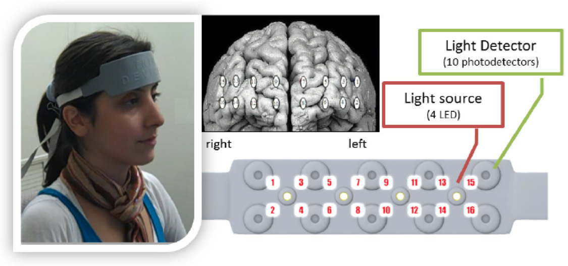
The image above depicts the fNIRS ‘headband’, which is lined with sensors. The headband is placed along the forehead and its sensors detect changes in brain activity.
[Image from Ayaz, H., Crawford, P., Curtin, A., Syed, M., Onaral, B., Beltman, W. M., & Shewokis, P. A. (2013, July). Differential prefrontal response during natural and synthetic speech perception: An fNIR based neuroergonomics study. In International Conference on Augmented Cognition (pp. 241-249). Springer, Berlin, Heidelberg].
WHAT DID THIS STUDY FIND?
- fNIRS detects THC-induced brain changes in the prefrontal cortex.
Of the 20 specific regions investigated in the prefrontal cortex, blood flow in three regions increased post-THC (all within the right orbitofrontal cortex). However, these brain changes were not significantly correlated (i.e. associated) with working memory task performance.
- Individuals reporting greater intoxication showed larger brain changes in the prefrontal cortex.
When participants were divided into high-intoxication and low-intoxication groups, the high- intoxication group exhibited more change in bloodflow from pre-THC to post-THC imaging sessions. More specifically, the high-intoxication group showed greater increases in bloodflow in three regions within the right and middle prefrontal cortex.
- In this small sample, working memory performance was not significantly impaired after THC.
Participants made slightly more errors on the working memory task post-THC (3.83 average errors made in the pre-THC session vs. 5.25 average errors made in the post-THC session). However, pre-THC and post-THC errors did not significantly differ from one another in statistical analyses. Similarly, reaction times on the working memory task were somewhat slower post-THC, but did not significantly differ from pre-THC measures. Nonsignificant findings are likely due to the small sample size of this study.
WHAT ARE THE IMPLICATIONS OF THE STUDY FINDINGS?
As the first fNIRS investigation of THC’s acute effects, this study suggests that this portable, non-invasive imaging technique can be used to investigate blood flow changes during cannabis intoxication. Given that individuals who reported higher intoxication showed the greatest changes in neural blood flow, this technique has the potential to detect THC intoxication in real time. Evaluation in real time has the potential to significantly benefit traffic safety. THC is shown to affect driving such that drivers may slow down in an attempt to compensate for intoxication, but skills suffer further as the situation becomes more demanding. Unlike alcohol, law enforcement does not currently have a reliable way of measuring THC intoxication in the field (e.g., automotive incidents where intoxication is suspected).
fNIRS is inexpensive, portable, and could pose a promising new technique for obtaining a more reliable measure of acute THC intoxication in non-clinical settings. Although working memory performance was not significantly impaired after THC administration, increased activity in the prefrontal cortex post-THC might suggest that the brain requires more neural resources (i.e. has to work harder) to perform the task when THC is on-board. Despite working memory performance not being related to blood flow changes in statistical tests, this study’s sample size was particularly small and further investigation with larger samples could reveal a significant relationship.
On the other hand, the THC-induced increase in prefrontal cortex blood flow may have effects on other cognitive functions not tested here. Brain regions that showed changes post-THC are associated with decision making skills and these structures are commonly implicated in addiction and impaired with other substance use. If these regions are generally affected by acute substance use, their THC-induced changes may have detremental effects on other cognitive functions. Further investigation is needed to determine the extent to which these THC-induced neural changes correspond to behavioral change.
- LIMITATIONS
-
- Without significant behavioral impairment, it is unclear what the impact of this abnormal frontal lobe activity is. Further investigation is needed to determine how THC-induced increased blood flow during cognitive task performance affects behavior. In this study, behavioral data was only available for 12 participants and a single task was used to test working memory skills. Perhaps investigation of a larger study sample with more difficult tasks or tasks that demand different cognitive functions, but elicit similar brain regions, would shed some light on the behavioral consequences of these frontal brain alterations.
- THC doses differed between participants. Perhaps a less variable and larger dose that depended less on participants’ self-reported history of cannabis use would result in more pronounced behavioral effects.
- The study sample primarily consisted of men and investigation did not include a control group. Placebo controlled studies of both men and women are needed to advance this area of study.
BOTTOM LINE
- For individuals & families seeking recovery: Given the widespread and growing use of cannabis in the U.S., it is important to understand THC’s effects on the brain and behavior, and to identify practical ways for measuring intoxication-related impairment, particularly as it relates to driving. This study is the first to use fNIRS to assess THC’s effects, suggesting that this inexpensive and easy-to-use imaging technique may help us to measure THC intoxication, similar to how we measure alcohol intoxication. It can ultimately help law enforcement to identify brain patterns of intoxication and increase traffic safety. Although participants who felt higher intoxication showed the greatest THC-related brain changes, it is not yet clear how these changes might impact cognitive ability. More research is needed to determine how THC-related brain changes relate to behavior and whether greater feelings of intoxication translate to greater cognitive impairment.
- For scientists: Brain imaging techniques are often expensive and sometimes invasive. fNIRS is a promising new technique offering less expensive, noninvasive ways for measuring changes in cerebral blood flow. The current study provides preliminary data suggesting that fNIRS could be used as a marker of THC-induced prefrontal cortex alterations and subjective intoxication. However, this investigation failed to find significant THC-related cognitive impairment in their small study sample. The pattern of results here demands a fully powered double-blind controlled investigation of THC’s acute effects with larger samples, additional neuropsychological assessments, and additional measures of subjective/objective intoxication.
- For policy makers: There is a growing population of adult recreational cannabis users in the U.S. and cannabis has acute affects that can negatively affect the brain and cognition. Accordingly, there has been a recent increase in the number of non-fatal and fatal vehicle collisions involving cannabis. Whether or not acute intoxication is responsible for these collisions is unclear. Unlike alcohol, we do not yet have a reliable technique for measuring THC intoxication. Studies like this provide promising preliminary evidence that we may one day be able to conduct roadside assessments for THC intoxication. Like a “THC breathalyzer”, fNIRS could provide a relatively inexpensive, non-invase way for law enforcement officials to measure cannabis-related changes in the brain that reflect intoxication. However, the current study was the first to use fNIRS to measure acute THC effects and funding is needed to conduct larger studies of this kind before we can realize the full potential of this promising technique.
- For treatment professionals and treatment systems: Existing research suggests that THC intoxication affects the brain and behavior. Permitting the inexpensive and non-invasive assessment of neural blood flow, fNIRS could allow clinicians and law-enforcement officials to detect real-time brain activity indicative of acute THC intoxication, ultimately aiding clinicians in patient management and law enforcement in traffic safety. However, this research is still in its early stages and further study is needed to further understand what these brain changes mean with respect to cognitive function and subjective/objective intoxication.
CITATIONS
Keles, H. O., Radoman, M., Pachas, G. N., Evins, A. E., & Gilman, J. M. (2017). Using Functional Near-Infrared Spectroscopy to Measure Effects of Delta 9-Tetrahydrocannabinol on Prefrontal Activity and Working Memory in Cannabis Users. Frontiers in human neuroscience, 11, 488.